Biomedical


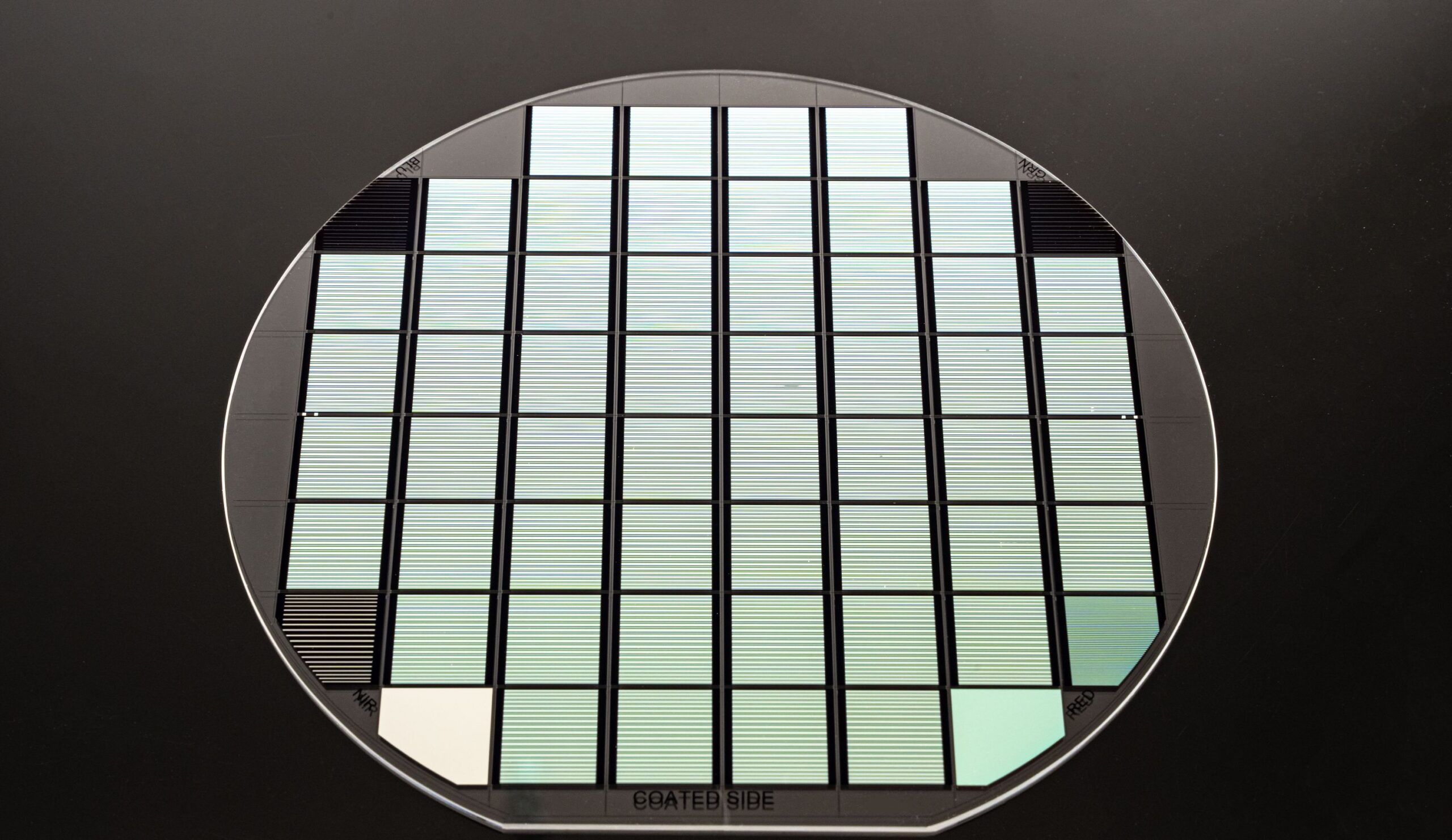
Biomedical experts use precise imaging details to make accurate diagnoses and innovative clinical advancements. Biomedical professionals rely on high-quality optical filters to get the most detailed images possible. Biomedical specialty filters provide the high-quality performance you need for your application.
Benefits of Our Biomedical Optical Filters
Our biomedical filters provide high transmission, creating bright images with deep blocking to ensure no unwanted light gets through. As a result, when you use our filters, your pictures will have a better signal-to-noise ratio than you’d get with many other standard filters.
We can also customize our filters to your exact specifications. In addition, our team can accommodate custom shapes, spectral specs, and other optics to achieve your desired imaging.
Fluorophores are fluorescent chemical compounds that absorb and emit light. They are often attached to molecules like peptides, proteins, amino acids, and antibodies and are used to label a specific target. These fluorescent probes are detected using a fluorescence-reading instrument, such as a plate reader or fluorescence microscope.
We provide filters that selectively transmit emitted wavelengths and block excitation wavelengths, improving contrast for both sensing and imaging fluorophores.
Fluorescence sensing and imaging have numerous biomedical applications, including:
- Labeling
- Chemical sensors
- Biological detectors
- Microscopy
- Immunofluorescence
- Tissue Sensing and Imaging
We provide custom filters that measure application-specific spectral bands for label-free and stained tissue sensing and imaging. As a result, you’ll get detailed insights into various tissue states, helping you more accurately analyze and diagnose the tissue.
Label-free tissue sensing and imaging eliminate the need for labels commonly required in techniques like fluorescence, providing technical, practical, and economic advantages for many applications. Various medical devices and instruments can evaluate tissue at different wavelengths to provide measurements and feedback on tissue characteristics like oxygenation levels.
Staining can also highlight structures in biological tissues — such as muscle fibers, cell populations, and cytological structures. Additionally, you can use staining to mark cells in flow cytometry or flag nucleic acids or proteins.
Clinical Imaging
Many clinical applications use Indocyanine green (ICG), a Food and Drug Administration (FDA)-approved fluorophore. ICG is used for:
- Blood flow visualization in the retina.
- Angiograms track blood flow in the heart.
- Blood vessel imaging during or after surgery.
- Perfusion after an amputation or transplant.
- Hepatic function studies.
- Wound healing.
We produce filters that isolate ICG by filtering out scattered light from an excitation beam to make clear, precise images.